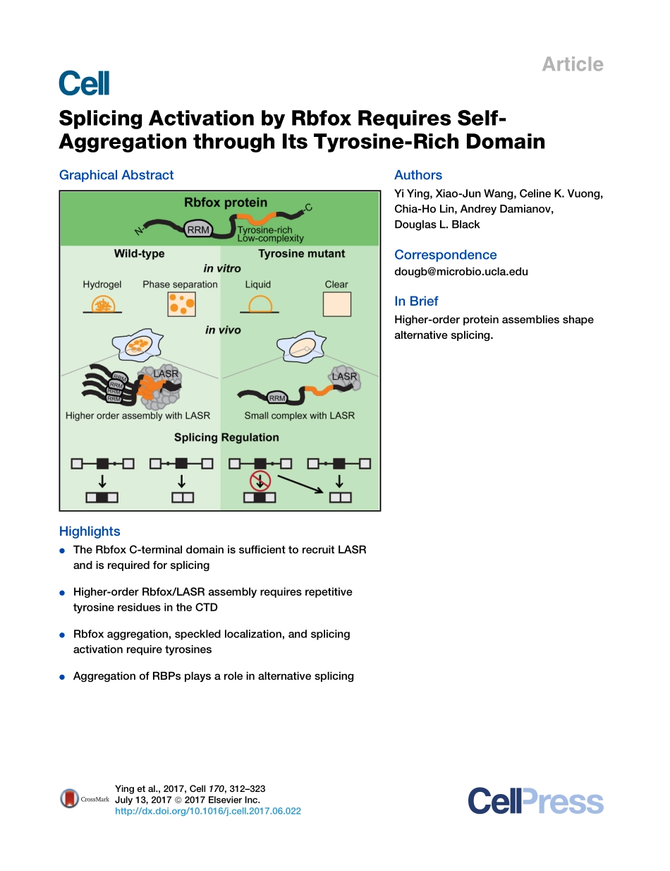
分享
2017-Ying-Splicing Activation by Rbfox Require.pdf
下载文档

ID:3519160

大小:3.73MB

页数:23页

格式:PDF

时间:2024-05-18

收藏 分享赚钱
温馨提示:
1. 部分包含数学公式或PPT动画的文件,查看预览时可能会显示错乱或异常,文件下载后无此问题,请放心下载。
2. 本文档由用户上传,版权归属用户,汇文网负责整理代发布。如果您对本文档版权有争议请及时联系客服。
3. 下载前请仔细阅读文档内容,确认文档内容符合您的需求后进行下载,若出现内容与标题不符可向本站投诉处理。
4. 下载文档时可能由于网络波动等原因无法下载或下载错误,付费完成后未能成功下载的用户请联系客服处理。
网站客服:3074922707
2017-Ying-Splicing Activation by Rbfox Require 2017 Ying Splicing
ArticleSplicing Activation by Rbfox Requires Self-Aggregation through Its Tyrosine-Rich DomainGraphical AbstractHighlightsdThe Rbfox C-terminal domain is sufficient to recruit LASRand is required for splicingdHigher-order Rbfox/LASR assembly requires repetitivetyrosine residues in the CTDdRbfox aggregation,speckled localization,and splicingactivation require tyrosinesdAggregation of RBPs plays a role in alternative splicingAuthorsYi Ying,Xiao-Jun Wang,Celine K.Vuong,Chia-Ho Lin,Andrey Damianov,Douglas L.BlackCorrespondencedougbmicrobio.ucla.eduIn BriefHigher-order protein assemblies shapealternative splicing.Ying et al.,2017,Cell 170,312323July 13,2017 2017 Elsevier Inc.http:/dx.doi.org/10.1016/j.cell.2017.06.022ArticleSplicing Activation by Rbfox RequiresSelf-Aggregation through Its Tyrosine-Rich DomainYi Ying,1,3Xiao-Jun Wang,2Celine K.Vuong,1,3Chia-Ho Lin,2Andrey Damianov,2and Douglas L.Black2,3,4,*1Molecular Biology Interdepartmental Doctoral Program2Department of Microbiology,Immunology,and Molecular Genetics3Molecular Biology InstituteUniversity of California,Los Angeles,Los Angeles,CA,USA4Lead Contact*Correspondence:dougbmicrobio.ucla.eduhttp:/dx.doi.org/10.1016/j.cell.2017.06.022SUMMARYProteins of the Rbfox family act with a complexof proteins called the Large Assembly of SplicingRegulators(LASR).We find that Rbfox interactswith LASR via its C-terminal domain(CTD),andthis domain is essential for its splicing activity.Inaddition to LASR recruitment,a low-complexity(LC)sequence within the CTD contains repeatedtyrosines that mediate higher-order assembly ofRbfox/LASR and are required for splicing activationby Rbfox.This sequence spontaneously aggregatesin solution to form fibrous structures and hydrogels,suggesting an assembly similar to the insolublecellular inclusions formed by FUS and other proteinsin neurologic disease.Unlike the pathological aggre-gates,we find that assembly of the Rbfox CTD playsan essential role in its normal splicing function.Rather than simple recruitment of individual regula-tors to a target exon,alternative splicing choicesalso depend on the higher-order assembly of theseregulators within the nucleus.INTRODUCTIONRNA-binding proteins(RBPs)control all aspects of RNA meta-bolism from biogenesis to decay(Singh et al.,2015).These pro-teins contain various classes of RNA-binding domains(RBDs)that recognize a wide range of short RNA elements,as well asauxiliary non-RNA binding domains that often contain intrinsi-cally disordered regions(IDRs)and sequences of low aminoacid complexity(LC)(Calabretta and Richard,2015).Recentstudies have found that certain IDR and LC domains can formfibers or phase-separated liquid droplets in vitro(Courchaineet al.,2016;Lin et al.,2015;Molliex et al.,2015;Nott et al.,2015;Schwartz et al.,2015).These sequences can furthercondense into highly stable hydrogel assemblies containing anamyloid-like cross-beta structure(Eisenberg and Jucker,2012;Kato et al.,2012;Murakami et al.,2015;Patel et al.,2015).Considerable interest in these aggregation properties stemsfrom the discovery that RBPs such as FUS and TARDBP formstableamyloid-likecellularinclusionsinamyotrophiclateralscle-rosis(ALS)and other neurological pathologies(Lagier-TourenneandCleveland,2009).Recentwork hasalso foundthatIDRorLCsequences function in normal cytoplasmic mRNA metabolism toallow the reversible concentration of RBPs within membrane-free subcellular organelles such as stress granules or ribonu-cleoprotein(RNP)granules(Courchaine et al.,2016;Jain et al.,2016;Kato et al.,2012;Kroschwald et al.,2015;Wallace et al.,2015).The nuclear RBPs involved in splicing also have extensiveLC sequences,but roles for these sequences and their aggrega-tion are not yet defined.Theregulationofalternativepre-mRNAsplicinginvolvesaverylarge number of RBPs that bind nascent transcripts to alter spli-ceosome assembly and splice site choice(Fu and Ares,2014;Lee and Rio,2015).One family of splicing regulators is the Rbfoxproteins that control networks of spliced isoform expression inbrain,heart,and muscle and during embryonic development(Conboy,2017).There are three mammalian Rbfox genes(RBFOX1,RBFOX2,andRBFOX3)eachwithasingleRNArecog-nition motif(RRM)that binds the short element(U)GCAUG(Auw-eter et al.,2006).Alternative promoters and alternative splicingdiversify the products of each gene,generating both nuclearand cytoplasmic isoforms(Damianov and Black,2010;Leeet al.,2009;Nakahata and Kawamoto,2005).The N-and C-ter-minal domains have segments of low amino acid complexity andundefined structure whose function is unknown(Figure S1A;data not shown).Clinical interest in the Rbfox proteins stems from findings thatRBFOX1 can be mutated in patients with autism spectrum disor-ders and epilepsy(Bill et al.,2013;Lal et al.,2013).Similarly,ho-mozygous null Rbfox1 mutations in the mouse brain lead to aseizure phenotype(Gehman et al.,2011).Changes in Rbfox1expression,and in the splicing and expression of Rbfox1 targettranscripts,were also observed in brains of Autism SpectrumDisorder(ASD)patients(Lee et al.,2016;Parikshak et al.,2016;Voineagu et al.,2011;Weyn-Vanhentenryck et al.,2014).Recently,we showed that Rbfox proteins regulate splicingwhile bound with the Large Assembly of Splicing Regulators(LASR),a multiprotein complex of eight RBPs(Damianov et al.,2016).Rbfox/LASR complexes sedimented at?55S on densitygradients,indicating assemblies larger than a single Rbfoxbound to a single LASR,but the nature and significance ofthe interactions leading to this higher-order assembly were312Cell 170,312323,July 13,2017 2017 Elsevier Inc.unresolved.Here,we report that the C-terminal domain(CTD)ofRbfox interacts with LASR and is required for splicing regulation.AnLCsequencewithintheCTDmediateshigher-orderassemblyof Rbfox/LASR and nuclear speckle localization in vivo,as wellas formation of fibrous aggregates and hydrogels in vitro.Muta-tions that block the higher-order assembly of Rbfox but not itsLASR interaction prevent proper splicing regulation and estab-lish a link between the biophysical properties of Rbfox aggrega-tion and its function in splicing.RESULTSThe CTD of Rbfox Mediates Interaction with LASR andHigher-Order AssemblyTo identify regions of Rbfox1 responsible for interacting withLASR,we generated Flp-In T-REx 293 cell lines stably express-ing deletion mutants of Rbfox1 tagged with the HA-FLAG epi-topes and the SV40 NLS(Figure 1A).We isolated Rbfox/LASRcomplexes from these cells by FLAG immunoprecipitation andpeptide elution,as previously described(Damianov et al.,2016).The complete set of LASR proteins was co-immunopre-cipitated with Rbfox missing either the N-terminal domain(NTD)or the RBD,indicating that these two regions are notessential for interaction with LASR(Figure 1B).Although allLASR components were present in these samples,some sub-units,such as hnRNP UL2,were isolated in lower amounts,sug-gesting that the NTD and RBD may provide additional contactsfor particular proteins.In contrast,deletion of the CTD(DCTD)abolished the interaction with LASR,and the CTD fragmentalone co-immunoprecipitated the LASR subunits with similar ef-ficiencytofull-length Rbfox1(Figure1B).Thus,theCTDprovidesthe primary contact with LASR.We previously found that Rbfox1 sediments in glycerol gradi-entsasalargerassemblythanpredictedforasingleRbfox/LASRcomplex(Damianov et al.,2016).Examining the mutant proteins,we found that the CTD fragment sedimented as a higher-ordercomplex of approximately 55S similar to the full-length protein,while DCTD remained at the top of the glycerol gradient(Figure 1C).These results demonstrate that the Rbfox CTD isnecessary and sufficient both to interact with LASR and toform higher-order complexes.Repetitive Tyrosine Residues within the CTD AreEssential for Higher-Order Assembly of Rbfox1Rbfox1,Rbfox2,and Rbfox3 paralogs all form the higher-ordercomplexes seen in gradients.These were observed both withthe endogenous proteins in mouse brain and with ectopically ex-pressedproteinsinHEK293cells(Damianovetal.,2016).Rbfox1and Rbfox2 also have muscle-specific variants derived fromthe inclusion of exon M43 instead of exon B40(Damianov andBlack,2010).Exons M43 and B40 encode related but not iden-tical amino acid sequences within the CTD(Figure 1D).Exam-ining the M43 variants on glycerol gradients,we found thatRbfox1_M43 peaked in the 55S region similar to Rbfox1_B40and Rbfox2_B40.Strikingly,Rbfox2_M43 did not form higher-order complexes but instead sedimented as a much smallerspecies near the top of the gradient(Figure 1D).The differencein sedimentation is attributed to the M43 segment,as ahybrid Rbfox1 protein containing the M43 exon of Rbfox2(Rbfox1_2M43)also largely sedimented as small complexes(Figure 1D).These data suggest that Rbfox2_M43 might lackresidues needed for the higher-order assembly.Aligning theexon sequences,it was notable that of three tyrosine residuesin the B40 exons,two were conserved in Rbfox1_M43 but allwere missing from Rbfox2_M43(Figure 1D).Looking at the CTD region,we found additional tyrosine resi-dues upstream and particularly downstream of exon B40,withthe downstream residues more closely spaced(Figure 1E).Toexamine the role of these tyrosines,we created a series ofmutant Rbfox1 proteins with increasing numbers of tyrosineschanged to serines or alanines.An Rbfox1 mutant with the threetyrosine residues of exon B40 replaced by serine or alanine sedi-mented only partially at 55S,with a substantial fraction of theprotein shifted to near the top of the gradient(Figure 1F,chang-ing tyrosines 13 in Figure 1E).Mutation of additional tyrosinesnearly eliminated higher-order assembly(Figure 1F,changingtyrosines 16,17,or 110 in Figure 1E).Serine substitutionsshowed a slightly stronger effect than alanine.In contrast,changing three tyrosines to phenylalanines did not impair thehigher-order assembly of Rbfox1,suggesting that aromatic in-teractions contribute to the assembly(Figure 1F).Given that the CTD was required for the Rbfox interaction withLASR,it was possible that the higher-order assembly involvedinteractions of LASR proteins and that the effect of the tyrosinemutations reflected a loss of LASR binding to Rbfox.This provednot to be the case.All the tyrosine-to-serine mutants as well asRbfox2_M43 retain their interaction with LASR,immunoprecipi-tating LASR with equal efficiency to wild-type protein(Figure 1G;data not shown).The property of higher-order assembly byRbfox is apparently separate from the LASR interaction.To further define the interactions between Rbfox and LASR,we divided the CTD region into three fragments,C1,C2,andC3(Figure 1E).C1 included the sequence upstream of the tenmutated tyrosines.C2 contained exon B40 and all ten of themutated tyrosines.C3 contained the extreme C terminusincluding some additional tyrosines and the NLS.Each of theseC-terminal regionswasfusedto theRbfox1DCTDprotein,whichdid not interact with LASR on its own(Figure 1B).Adding C1 toRbfox1 DCTD had little effect,with the protein pulling downonlysmallamountsof LASR(Figure 2A,bottom),andnotforminghigher-order complexes(Figure 2B).In contrast,Rbfox1 DCTDfused to either C2 or C3 pulled down LASR relatively efficiently(Figure 2A,bottom).C2 was more prone to aggregation intohigher-order complexes than C3(Figure 2B).The medium-sizecomplexes in fractions 79 formed by C3 may result from tyro-sines not tested by mutation.Mutation of the tyrosines in C2 toserines eliminated the higher-order assembly and unlike thefull-length protein also reduced the interaction with LASR(Fig-ure 2).These data indicate that subregions of the CTD can inde-pendently bind LASR and that in some segments the tyrosinescontribute to this interaction.Aggregation of the CTD AloneAvarietyofaggregationpropertieshavebeenreportedfortheLCsequences of RBPs,including the formation of hydrogels,amy-loid-like fibrils,and phase-separated liquid droplets(Aguzzi andCell 170,312323,July 13,2017313ABCDFEGFigure 1.The Rbfox CTD Mediates Both LASR Binding and Higher-Order Assembly(A)Diagram of Rbfox1 domains and deletion mutants.(B)Co-immunoprecipitation ofLASRwithRbfox1andmutants.Solubleandhigh-molecular-weight(HMW)nuclearfractionswerepreparedfromFlp-InT-REx293cells stably expressing either full-length HA-FLAG-SV40NLS-Rbfox1(FL)or a deletion mutant as diagrammed in(A).Anti-FLAG immunoprecipitates were elutedwith FLAG peptide,separated on gels,and stained with SYPRO Ruby.Arrowheads indicate HA-FLAG-SV40NLS-Rbfox1 protein and its mutants.LASR subunitsare indicated on the right.(C)Sedimentation of Rbfox1 complexes in glycerol density gradients.HMW fractions of cells expressing Rbfox1 or an Rbfox1 fragment were loaded onto 10%50%glycerol density gradients.Gradient fractions from top to bottom run from left to right.Proteins from odd fractions were immunoblotted with anti-FLAGantibody.40S and 60S markers are indicated below.(D)Amino acid sequences encoded by exons B40 and M43 in Rbfox1 and Rbfox2(top).Sedimentation of Rbfox1 and Rbfox2 splice variants in glycerol densitygradients as in(C)(bottom).(E)CTD sequence alignments for the nuclear B40 isoforms of Rbfox1,Rbfox2,and Rbfox3.Tyrosines examined by mutagenesis are shown in red.Sequences ofC1,C2,and C3 fragments of CTD are indicated by arrows.(F)Sedimentation of Rbfox1 tyrosine mutants in glycerol density gradients.Rbfox1 proteins containing increasing numbers of mutated tyrosines were separatedas in(C).Three to ten tyrosine residues were mutated to serine,alanine,or phenylalanine as indicated on the left.(G)Immunoprecipitation of LASR with HA-FLAG-Rbfox1 containing tyrosine-to-serine mutations.Rbfox proteins and LASR subunits are indicated on the right.See also Figure S1.314Cell 170,312323,July 13,2017Altmeyer,2016;Bergeron-Sandoval et al.,2016;Kato et al.,2012;Lin et al.,2015).The LC sequence of the FUS protein con-tains the repeated tripeptide G/SYG/S that mediates its as-sembly into an amyloid-like cross-beta structure(Kato et al.,2012).Although not exact matches to the FUS tripeptide motif,many of the tyrosines in the Rbfox1 C2 region are preceded orfollowed by glycine or serine.We found that polypeptides ofthe CTD were predicted as potential hotspots of amyloidal ag-gregation by AGGRESCAN(Conchillo-Sole et al.,2007)(Fig-ure S1A).To examine the aggregation properties of the RbfoxCTD,we purified His-tagged recombinant proteins containingthe wild-type CTD or its mutant with ten tyrosines changed toserines(CTD-YS),each fused to a SNAP tag(Figure S1D).TheSNAP-CTD fusion eluted largely in the void volume from asize exclusion column,with some unaggregated monomer.Incontrast,nearly all of the CTD-YS mutant eluted as monomericprotein,indicating that it is less prone to aggregation(Fig-ure S1B).We also made equivalent fusions with only the C2 frag-ment and its YS mutant.The SNAP-C2 fusion presented elutionpeaks in the void,as a multimer of about 150 kDa,and as mono-mer(Figure S1B).For the SNAP-C2-YS mutant,the void volumepeak

此文档下载收益归作者所有

下载文档
收起
展开