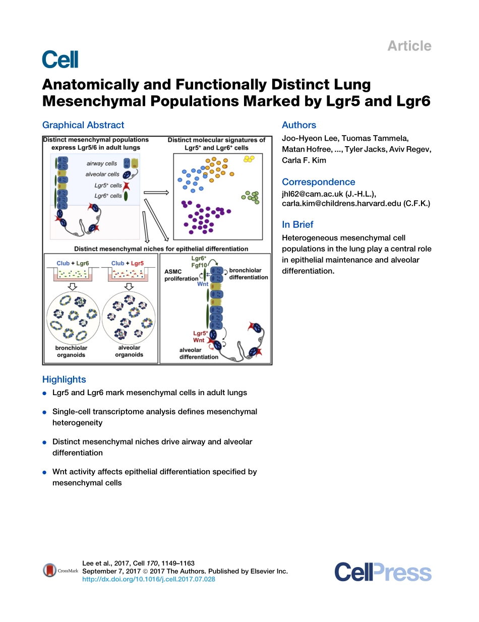
分享
2017-Lee-Anatomically and Functionally Distinc.pdf
下载文档

ID:3519157

大小:10.21MB

页数:28页

格式:PDF

时间:2024-05-18

收藏 分享赚钱
温馨提示:
1. 部分包含数学公式或PPT动画的文件,查看预览时可能会显示错乱或异常,文件下载后无此问题,请放心下载。
2. 本文档由用户上传,版权归属用户,汇文网负责整理代发布。如果您对本文档版权有争议请及时联系客服。
3. 下载前请仔细阅读文档内容,确认文档内容符合您的需求后进行下载,若出现内容与标题不符可向本站投诉处理。
4. 下载文档时可能由于网络波动等原因无法下载或下载错误,付费完成后未能成功下载的用户请联系客服处理。
网站客服:3074922707
2017-Lee-Anatomically and Functionally Distinc 2017 Lee Anatomically
ArticleAnatomically and Functionally Distinct LungMesenchymal Populations Marked by Lgr5 and Lgr6Graphical AbstractHighlightsdLgr5 and Lgr6 mark mesenchymal cells in adult lungsdSingle-cell transcriptome analysis defines mesenchymalheterogeneitydDistinct mesenchymal niches drive airway and alveolardifferentiationdWnt activity affects epithelial differentiation specified bymesenchymal cellsAuthorsJoo-Hyeon Lee,Tuomas Tammela,MatanHofree,.,TylerJacks,AvivRegev,Carla F.KimCorrespondencejhl62cam.ac.uk(J.-H.L.),carla.kimchildrens.harvard.edu(C.F.K.)In BriefHeterogeneous mesenchymal cellpopulations in the lung play a central rolein epithelial maintenance and alveolardifferentiation.Lee et al.,2017,Cell 170,11491163September 7,2017 2017 The Authors.Published by Elsevier Inc.http:/dx.doi.org/10.1016/j.cell.2017.07.028ArticleAnatomically and Functionally Distinct LungMesenchymal Populations Marked by Lgr5 and Lgr6Joo-Hyeon Lee,1,2,3,4,5,*Tuomas Tammela,6Matan Hofree,7Jinwook Choi,4Nemanja Despot Marjanovic,6Seungmin Han,4,8David Canner,6Katherine Wu,6Margherita Paschini,1,2,3Dong Ha Bhang,9Tyler Jacks,6,10Aviv Regev,6,7,10and Carla F.Kim1,2,3,11,*1Stem Cell Program and Divisions of Hematology/Oncology and Pulmonary&Respiratory Diseases,Boston Childrens Hospital,Boston,MA02115,USA2Harvard Stem Cell Institute,Cambridge,MA 02138,USA3Department of Genetics,Harvard Medical School,Boston,MA 02115,USA4Wellcome Trust/Medical Research Council Stem Cell Institute,University of Cambridge,Tennis Court Road,Cambridge CB2 1QR,UK5Department of Physiology,Development and Neuroscience,University of Cambridge,Cambridge CB2 3DY,UK6David H.Koch Institute for Integrative Cancer Research,Massachusetts Institute of Technology,Cambridge,MA 02142,USA7Broad Institute of MIT and Harvard,Cambridge,MA 02142,USA8Wellcome Trust/Cancer Research UK Gurdon Institute,University of Cambridge,Tennis Court Road,Cambridge CB2 1QN,UK9DepartmentofCancerBiology,AbramsonFamilyCancerResearchInstitute,UniversityofPennsylvaniaSchoolofMedicine,Philadelphia,PA19104,USA10Howard Hughes Medical Institute,Department of Biology,Massachusetts Institute of Technology,Cambridge,MA 02139,USA11Lead Contact*Correspondence:jhl62cam.ac.uk(J.-H.L.),carla.kimchildrens.harvard.edu(C.F.K.)http:/dx.doi.org/10.1016/j.cell.2017.07.028SUMMARYThe diversity of mesenchymal cell types in the lungthat influence epithelial homeostasis and regenera-tion is poorly defined.We used genetic lineagetracing,single-cell RNA sequencing,and organoidculture approaches to show that Lgr5 and Lgr6,well-known markers of stem cells in epithelialtissues,are markers of mesenchymal cells in theadult lung.Lgr6+cells comprise a subpopulation ofsmooth muscle cells surrounding airway epitheliaand promote airway differentiation of epithelialprogenitors via Wnt-Fgf10 cooperation.Geneticablation of Lgr6+cells impairs airway injury repairin vivo.Distinct Lgr5+cells are located in alveolarcompartments and are sufficient to promote alveolardifferentiation of epithelial progenitors through Wntactivation.Modulating Wnt activity altered differenti-ation outcomes specified by mesenchymal cells.This identification of region-and lineage-specificcrosstalk between epithelium and their neighboringmesenchymal partners provides new understandingof how different cell types are maintained in the adultlung.INTRODUCTIONHomeostasis and injury repair of the adult lung epithelium involvethe active engagement of epithelial cell populations that reside indistinctanatomicallocations.Inthedistallung,multipleprogenitorpopulationshavebeenshowntoparticipateintherepairprocess.The different anatomical locations of diverse epithelial progenitorcellsinthelungmake itlikely thatdistinctstromalfactorsregulatethe behavior of these cells.However,understanding the precisemolecular mechanisms influencing progenitor cells is precludedby limited knowledge of stromal cell identities in the lung.Defining the identities and behavior of lung mesenchymal cellsis challenging due to the lack of defined markers for these pop-ulations.During lung development,the mesenchymal progeni-tors undergo regionally distinct differentiation programs,givingrise to airway and vascular smooth muscle,alveolar fibroblasts,endothelium,and pericytes,among others(McCulley et al.,2015).Clonal analysis illustrated the diversity of mesenchymalprogenitors(Kumar et al.,2014).Mesenchyme expressing fibro-blast growth factor 10(Fgf10),glioma-associated oncogene1(Gli1),and Axin2 contribute to smooth muscle and alveolarfibroblast-like cells(El Agha et al.,2014;Li et al.,2015;Mailleuxetal.,2005;Moiseenkoetal.,2017).However,theinformation onthespatialheterogeneityandbehaviorofmesenchymalcellsthatimpact epithelial progenitors in lung regeneration and repairremains unclear.In the airway epithelium of the adult murine distal lungs,clubcells(formerly known as Clara cells)function as progenitorsthat can both self-renew and produce differentiated ciliated cellsat steady state(Rawlins et al.,2009).Following airway injuryusing naphthalene,which abolishes Cyp2f2-expressing clubcells,the surviving club cells can divide and regenerate theairwayepithelium(Hoganetal.,2014).Lineage-tracingapproaches showed that cells expressing the club cell markerCCSP,encoded bythe Scgb1a1 gene,are alsocapable of givingrise to alveolar lineage cells following bleomycin-induced alve-olar damage(Rock et al.,2011).However,little is known aboutthe precise mechanisms regulating club cell behavior duringrepair and regenerative processes.Wnt signals function in development and regeneration of thelung(Cardoso and Lu,2006;Hogan et al.,2014),whereas littleCell 170,11491163,September 7,2017 2017 The Authors.Published by Elsevier Inc.1149This is an open access article under the CC BY license(http:/creativecommons.org/licenses/by/4.0/).Wnt activity is documented in the normal adult lung.Recentstudies have uncovered a small family of 7-transmembranereceptors,leucine-rich repeat-containing G protein-coupledreceptor-5(Lgr5)family,comprising Lgr4,Lgr5,and Lgr6(Clevers et al.,2014).Lgr5 is specifically expressed in epithelialstem cells in multiple tissues,including the intestine,liver,andskin(Barker et al.,2007,2010;Huch et al.,2013;Jaks et al.,2008).Lgr6 expression has been reported in bipotent skin pro-genitor cells(Snippert et al.,2010).More recently,Wnt-respon-sivecellsexpressingLgr5werereportedtobehighlyproliferativeand progressive in lung adenocarcinoma(Tammela et al.,2017).Here,we used single-cell RNA sequencing(scRNA-seq),line-age tracing,and organoid cultures to characterize adult lungmesenchymal populations marked by Lgr5 and Lgr6.Lgr6-ex-pressing cells were found surrounding bronchiolar epitheliaand in the alveolar space,whereas Lgr5-expressing cells werelargely alveolar.Ex vivo organoid co-culture of Scgb1a1 line-age-labeled cells with Lgr6-expressing cells revealed theLgr6+cellsdirect airwaydifferentiation ofScgb1a1+progenitors.In contrast,Lgr5-expressing mesenchymal cells promote alve-olar differentiation via activation of Wnt pathway.These resultsdemonstrate that region-specific crosstalk between airwaystem cells and adjacent mesenchymal cells is required to main-tain proper tissue integrity.RESULTSLgr5 and Lgr6 Mark Distinct Mesenchymal CellPopulations in Adult LungTo investigate the functional role of Lgr5 and Lgr6 in adult lungs,we characterized Lgr6 expression in the lung using Lgr6-EGFP-IRES-CreERT2 knockin mice,in which EGFP marks cells withactive expression of the Lgr6 locus(Snippert et al.,2010).Unex-pectedly,rather than marking epithelial cells,Lgr6-expressingcells were found throughout the lung mesenchyme surroundingthe conducting airways.Immunohistochemistry showed thatthese cells express a-smooth muscle actin(a-SMA)(encodedby Acta2),a marker of smooth muscle cells(Figure 1A).Notably,no Lgr6 expression was observed in vascular smooth musclecells(VSMCs)(arrowhead,Figure 1A).In the alveolar regions,we found scattered EGFP-positive cells(GFP+)that are negativefor a-SMA(Figure 1B).Fluorescence-activated cell sorting(FACS)analysisrevealedthat9.12%1.42%ofresidentmesen-chymal cells(GFP+/CD31?CD45?EpCAM?)express Lgr6 inadult lungs(Figure 1C).qPCR confirmed that these populationsrobustly express Lgr6 and Acta2.Wealso detected Lgr5 expres-sion in the Lgr6+cells,suggesting Lgr6 may mark cell popula-tions expressing Lgr5(Figure 1D).We next utilized Lgr5-IRES-CreERT2 mice that were crossedto a Rosa26-lox-stop-lox-TdTomato reporter allele(hereafter,Lgr5-CreERT2;R26-Tom)to investigate expression of Lgr5 inadult lungs.In contrast with Lgr6+cells,the majority of lineage-labeled Lgr5+cells were located exclusively in the alveolar com-partments and none of the lineage-labeled cells were airwaysmooth muscle cells(ASMCs)(Figures 1E and 1F).A small num-berofcellsthatwerenegativefora-SMAwerefoundnearairways(Figure S1).FACS analysis indicated that 1.24%0.42%of resi-dent lung mesenchymal cells(Tom+/CD31?CD45?EpCAM?)were lineage-labeled by Lgr5(Figure 1G).In contrast to the highexpression level of Lgr5 in Lgr6+cells,expression of Lgr6 andActa2 was not highly enriched in the cell populations labeled byLgr5(Figures 1D and 1H).These results suggest that Lgr5 andLgr6 mark distinct mesenchymal lineages in adult lungs;the ma-jorityofLgr6+cellsareASMCs,whereasLgr5+cellsarefoundpri-marily in the alveolar regions.Heterogeneity of Mesenchymal Populations ExpressingLgr5 and Lgr6 in Adult LungsTo characterize the mesenchymal lineages labeled by Lgr5 andLgr6,we performed scRNA-seq of individual cells isolatedfrom Lgr5-CreERT2;R26-Tom and Lgr6-EGFP-CreERT2 mice(two replicates of each;Figure S2B).We purified single-cellsuspensions of dissociated Lgr5+and Lgr6+cells by FACSsorting with depletion of endothelial and immune cells(Lgr5,CD31?CD45?CD11b?TER119?Tom+;Lgr6,CD31?CD45?CD11b?TER119?GFP+;Figure 2A).We analyzed profiles from182 mesenchymal cells that passed strict quality-control thresh-olds(STAR Methods)and used a community detection clus-tering algorithm on k-nearest neighbor(k-NN)cell graph,createdfrom random subsamples of the data,to identify robust clustersby consensus clustering(STAR Methods).We identified five robust clusters of cell populations withdistinct transcriptional programs(clusters AE;Figures 2B2Dand S2B).Using marker genes known to be expressed in variousmesenchymalcells,wedistinguishedLgr5-andLgr6-expressingcell types that are regionally distributed.Specifically,clusterE cells had a distinctive high expression of Lgr6 and Acta2 butlow expression of Lgr5,suggesting this is the Lgr6-expressingASMCs population(Figures 2B2E,S2B,and S2C).Cells in thecluster also highly expressed various mesenchymal genes,such as Cspg4,Tagln,and Gli1.Clusters B and D cells exhibitedenriched expression of Lgr5.Some of the cells in these two clus-ters showed considerable Lgr6 expression,suggesting that thispopulation contains alveolar mesenchymal cells labeled by Lgr5and Lgr6(Figures 2B2E,S2B,and S2D).A distinct small popu-lation,clusterC,highlyexpressesgenesassociatedwithalveolarfibroblasts:Pdgfra;Wnt2;Fgf10;and Vcam1(Figures 2B2E,S2B,and S2E).Cells in all clusters expressing Lgr5 orLgr6 also expressed general mesenchymal markers,such asCol1a1,Vimentin,and Pdgfrb(Figure S2F).Cluster A,a distinctsmall cluster of cells,expressed the epithelial marker EpCAMand lung epithelial lineage markers,such as Scgb1a1(club cellmarker)and Sftpc(alveolar type II cell marker;Figures 2B2E,S2B,and S2G).Lineage-tracing studies confirmed that thereare rare cells expressing CCSP in lineage-labeled Lgr5+andLgr6+cells(Figures S2H and S2I).Each of the cell populationssuggested by cluster analysis expressed a unique gene expres-sion signature(Figures 2D and 2E).Taken together,scRNA-seqanalysis shows that the cellular heterogeneity of lung mesen-chymalcellsexpressingLgr5andLgr6isassociatedwithdistinctand separable transcriptional programs.Long-Term Tracing of Lgr6+Mesenchymal Cells in AdultLungs in HomeostasisTo evaluate the cellular behavior of Lgr6+cells in adult lungs,weestablished Lgr6-EGFP-CreERT2;R26-Tom mice.To determine1150Cell 170,11491163,September 7,2017whether lineage-labeled Lgr6+cells contribute to ASMC mainte-nance,we measured the proportion of lineage-labeled Lgr6+cells that are positive for a-SMA cells over time(Figure 3A).FACSanalysisshowedthatrecombinationoccurredin62.7%4.3%of Lgr6-expressing cells(Tom+GFP+/GFP+)at10 days after final Tmx injection(Figure 3B).As shown in Figures3C and 3D,the proportion of lineage-labeled Lgr6+a-SMA+ASMCs remained constant over the 12-month chase period.To further explore whether Lgr6+cells are resting cells orundergoproliferationinthesteadystate,Lgr6-EGFP-CreERT2;R26-Tom mice were injected with a single low doseof Tmx,which labels only a small proportion of Lgr6+cells(Figure 3F).Lungs were harvested over 12-month chase period,and sections were analyzed with confocal microscopy forthe presence of lineage-labeled cells or clusters that spanbothperi-airwayandalveolarcompartments(Figure3E).Figure 1.Distinct Mesenchymal Lineages Expressing Lgr5 and Lgr6 in Adult Lungs(A and B)Representative confocal images showing expression patterns of Lgr6 in adult distal lungs:GFP(green);a-SMA(yellow);and DAPI(blue)in lung tissuesections from Lgr6-EGFP-CreERT2 mice.Arrowheads indicate vascular smooth muscle cells expressing a-SMA+.aw,airway;v,blood vessel.(C)Representative profile of FACS-sorted EGFP+populations from Lgr6-EGFP-CreERT2 mice for qPCR analysis.(D)Validation of differential expression of Lgr5,Lgr6,and Acta2 in Lgr6+and Lgr6?cells by qPCR analysis.Expression from Lgr6+cells is shown as fold changerelative to Lgr6?cells set to 1,followed by normalization to Gapdh.(Eand F)Representativeconfocal images showing expression patterns of Lgr5 inadult distal lungs:Tdtomato(red);a-SMA(yellow);and DAPI(blue)inlung tissuesections from Lgr5-CreERT2;R26-Tom mice,followed by Tamoxifen injection.aw,airway;v,blood vessel.(G)Representative profile of FACS-sorted TdTomato+populations from Lgr5-CreERT2;R26-Tom mice for qPCR analysis.Sorting scheme is same as in(C).(H)Validation of differential expression of Lgr5,Lgr6,and Acta2 in Lgr5+and Lgr5?cells by qPCR analysis.Normalized as in(D).The scale bars represent 100 mm.Data presented are the mean of three independent experiments with triplicates.Error bars indicate SD(*p 0.001).See alsoFigure S1.Cell 170,11491163,September 7,20171151Single-labeled peri-airway or alveolar cells were widely distrib-uted at 10 days after Tmx administration(Figure 3G).Notably,after approximately 6 months,a few small clusters of lineage-labeled peri-airway cells were observed.Longer chases,up to12 months,confirmed that lineage-labeled cells are long livedand can be found in clusters,suggesting that there is a subsetof Lgr6+cells that are capable of proliferation with a slow rateat steady state.The size distribution of clusters became increas-ingly heterogeneous,but mean clone size and number of cellscomposed of clusters increased over tim

此文档下载收益归作者所有

下载文档
收起
展开