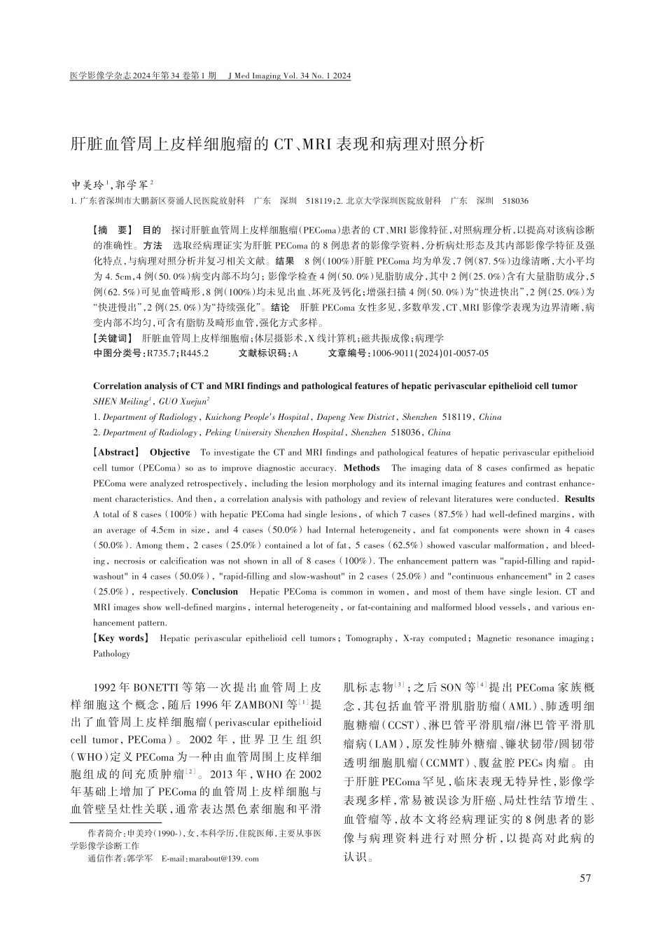
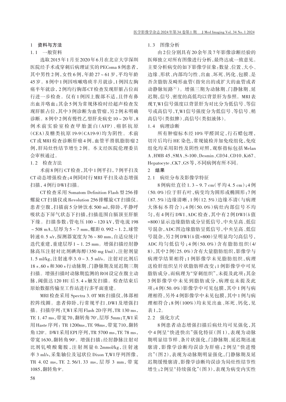
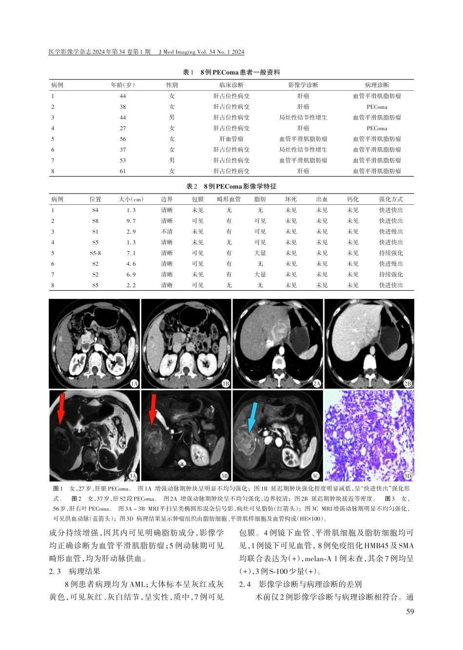
医学影像学杂志2024年第34卷第1期JMedImagingVol.34No.12024肝脏血管周上皮样细胞瘤的CT、MRI表现和病理对照分析申美玲1,郭学军21.广东省深圳市大鹏新区葵涌人民医院放射科广东深圳518119;2.北京大学深圳医院放射科广东深圳518036【摘要】目的探讨肝脏血管周上皮样细胞瘤(PEComa)患者的CT、MRI影像特征,对照病理分析,以提高对该病诊断的准确性。方法选取经病理证实为肝脏PEComa的8例患者的影像学资料,分析病灶形态及其内部影像学特征及强化特点,与病理对照分析并复习相关文献。结果8例(100%)肝脏PEComa均为单发,7例(87.5%)边缘清晰,大小平均为4.5cm,4例(50.0%)病变内部不均匀;影像学检查4例(50.0%)见脂肪成分,其中2例(25.0%)含有大量脂肪成分,5例(62.5%)可见血管畸形,8例(100%)均未见出血、坏死及钙化;增强扫描4例(50.0%)为“快进快出”,2例(25.0%)为“快进慢出”,2例(25.0%)为“持续强化”。结论肝脏PEComa女性多见,多数单发,CT、MRI影像学表现为边界清晰,病变内部不均匀,可含有脂肪及畸形血管,强化方式多样。【关键词】肝脏血管周上皮样细胞瘤;体层摄影术,X线计算机;磁共振成像;病理学中图分类号:R735.7;R445.2文献标识码:A文章编号:1006-9011(2024)01-0057-05CorrelationanalysisofCTandMRIfindingsandpathologicalfeaturesofhepaticperivascularepithelioidcelltumorSHENMeiling1,GUOXuejun21.DepartmentofRadiology,KuichongPeople'sHospital,DapengNewDistrict,Shenzhen518119,China2.DepartmentofRadiology,PekingUniversityShenzhenHospital,Shenzhen518036,China【Abstract】ObjectiveToinvestigatetheCTandMRIfindingsandpathologicalfeaturesofhepaticperivascularepithelioidcelltumor(PEComa)soastoimprovediagnosticaccuracy.MethodsTheimagingdataof8casesconfirmedashepaticPEComawereanalyzedretrospectively,includingthelesionmorphologyanditsinternalimagingfeaturesandcontrastenhance‐mentcharacteristics.Andthen,acorrelationanalysiswithpathologyandreviewofrelevantliteratureswereconducted.ResultsAtotalof8cases(100%)withhepaticPEComahadsinglelesions,ofwhich7cases(87.5%)hadwell-definedmargins,withanaverageof4.5cminsize,and4cases(50.0%)hadInternalheterogeneity,andfatcomponentswereshownin4cases(50.0%).Amongthem,2...