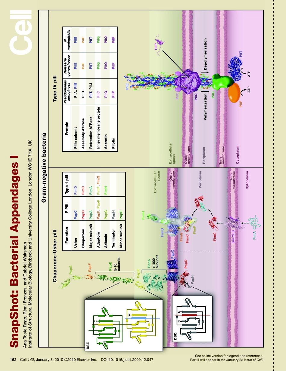
SnapShot:BacterialAppendagesIAnaTosteRego,RémiFronzes,andGabrielWaksmanInstituteofStructuralMolecularBiology,BirkbeckandUniversityCollegeLondon,LondonWC1E7HX,UKSeeonlineversionforlegendandreferences.PartIIwillappearintheJanuary22issueofCell.162Cell140,January8,2010©2010ElsevierInc.DOI10.1016/j.cell.2009.12.047Seeonlineversionforlegendandreferences.162.e1Cell140,January8,2010©2010ElsevierInc.DOI10.1016/j.cell.2009.12.047SnapShot:BacterialAppendagesIAnaTosteRego,RémiFronzes,andGabrielWaksmanInstituteofStructuralMolecularBiology,BirkbeckandUniversityCollegeLondon,LondonWC1E7HX,UKThistwo-partSnapShotdepictstheassemblyandstructureofselectednonflagellarsurfaceappendagesfromgram-negativebacteria.Theseincludechaperone-usherpiliandtypeIVpili(inpartI)andthetypeIIIsecretionneedleandtypeIVsecretionpili(inpartII).Chaperone-UsherPili(Left)Chaperone-usher(CU)pili,exemplifiedbythePandType1pili,arethemostabundantgroupofbacterialsurfaceappendages.Theyplayimportantrolesinadhesionandhostrecognition.Type1andPpiliarecomposedoftwomajorsubassemblies(seepanelontheleftforPpili):athickandrigidrodmadeupofsubunitsofPapAinPpiliorFimAinType1pili(lightblue)arrangedinaright-handedhelicalcylinderandathin,flexibletipfiberinthedistalendoftherod.InPpilithetipfiberiscomposedof5–10PapEsubunits(darkgreen)flankedbytwominorsubunits,PapF(orange)andPapK(yellow).InType1pilithelineartipfiberisformedbyonesubunitFimGandonesubunitFimF.Bothsystemshaveanadhesinsubunit(PapGandFimHinPandType1pili,respectively)atthetipofthefiber(lightgreen),responsibleforinteractionwiththehosttissues.InPpiliaterminationsubunitPapH(gray)anchorsthepilusstructuretothecellwall(WaksmanandHultgren,2009).Pilusassembly(seepanelontheright)isorchestratedbyaperiplasmicchaperonePapDforPpiliorFimCforType1pili(red),andanoutermembrane(OM)assemblyplatform,thedimericusherPapCforPpili,orFimDforType1pili(darkblue)(WaksmanandHultgren,2009).Pilussubunitscrosstheinnermembrane(IM)totheperiplasmviaSecYEG(pink).Thechaperonethencapturessubunitsviaamecha...