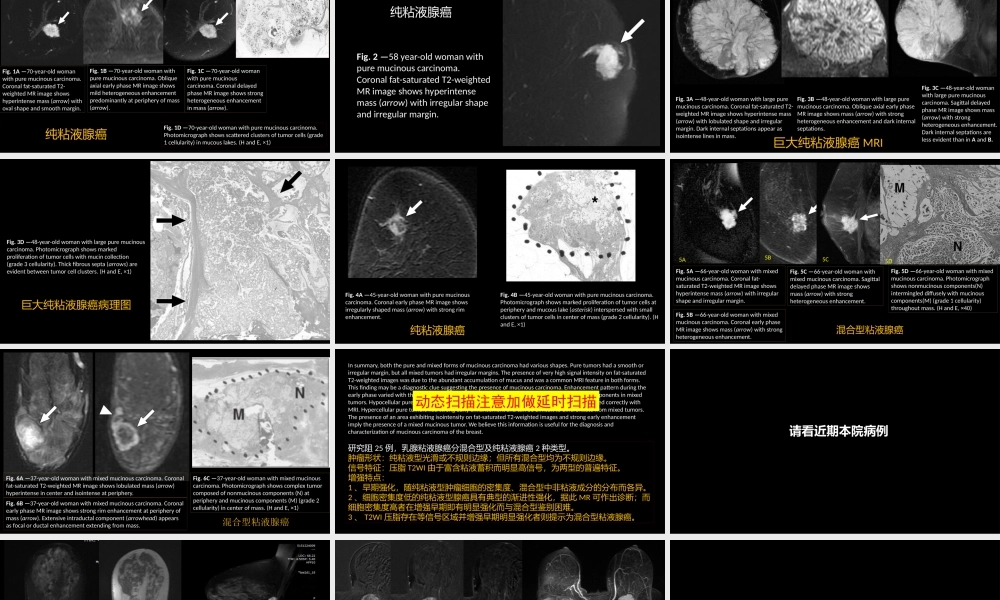
MucinousCarcinomaoftheBreast:MRIFeaturesofPureandMixedFormswithHistopathologicCorrelationShuichiMonzawaetalAJRMarch2009,VOLUME192NUMBER3乳腺粘液腺癌:纯粘液型、混合型粘液腺癌MRI特征与病理的关系杨景震编译Fig.1A—70-year-oldwomanwithpuremucinouscarcinoma.Coronalfat-saturatedT2-weightedMRimageshowshyperintensemass(arrow)withovalshapeandsmoothmargin.Fig.1B—70-year-oldwomanwithpuremucinouscarcinoma.ObliqueaxialearlyphaseMRimageshowsmildheterogeneousenhancementpredominantlyatperipheryofmass(arrow).Fig.1C—70-year-oldwomanwithpuremucinouscarcinoma.CoronaldelayedphaseMRimageshowsstrongheterogeneousenhancementinmass(arrow).Fig.1D—70-year-oldwomanwithpuremucinouscarcinoma.Photomicrographshowsscatteredclustersoftumorcells(grade1cellularity)inmucouslakes.(HandE,×1)纯粘液腺癌ABCDFig.2—58year-oldwomanwithpuremucinouscarcinoma.Coronalfat-saturatedT2-weightedMRimageshowshyperintensemass(arrow)withirregularshapeandirregularmargin.纯粘液腺癌Fig.3A—48-year-oldwomanwithlargepuremucinouscarcinoma.Coronalfat-saturatedT2-weightedMRimageshowshyperintensemass(arrow)withlobulatedshapeandirregularmargin.Darkinternalseptationsappearasisointenselinesinmass.Fig.3B—48-year-oldwomanwithlargepuremucinouscarcinoma.ObliqueaxialearlyphaseMRimageshowsmass(arrow)withstrongheterogeneousenhancementanddarkinternalseptations.Fig.3C—48-year-oldwomanwithlargepuremucinouscarcinoma.SagittaldelayedphaseMRimageshowsmass(arrow)withstrongheterogeneousenhancement.DarkinternalseptationsarelessevidentthaninAandB.巨大纯粘液腺癌MRIFig.3D—48-year-oldwomanwithlargepuremucinouscarcinoma.Photomicrographshowsmarkedproliferationoftumorcellswithmucincollection(grade3cellularity).Thickfibroussepta(arrows)areevidentbetweentumorcellclusters.(HandE,×1)巨大纯粘液腺癌病理图Fig.4A—45-year-oldwomanwithpuremucinouscarcinoma.CoronalearlyphaseMRimageshowsirregularlyshapedmass(arrow)withstrongrimenhancement.Fig.4B—45-year-oldwomanwithpuremucinouscarcinoma.Photomicrographshowsmarkedproliferationoftumorcellsatperipheryandmucou...